Effects of axial loaded magnetic resonance imaging of lumbar spine on dural sac and lateral recesses
DOI:
https://doi.org/10.17532/jhsci.2021.1589Keywords:
Spinal canal stenosis, axial loading, MRI, lumbar spine, dural sac cross-sectional area, depth lateral recessAbstract
Introduction: Axial-loaded magnetic resonance imaging (MRI), which can simulate an upright position of the patient may cause a significant reduction of the dural sac cross-sectional area (DCSA) compared with standard MRI, thus providing valuable information in the assessment of the lumbar spinal canal. The purpose of this study was to investigate excessiveness of the change in DCSA and depth of lateral recesses (DLRs) before and after axial-loaded imaging in relation to body mass index (BMI) of the subjects.
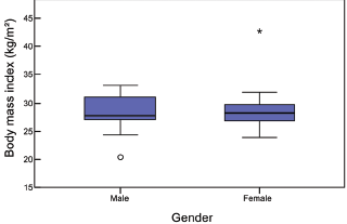
Methods: Twenty patients were scanned to evaluate DCSA and DLR at three consecutive lumbar spine intervertebral disc levels (L3/4, L4/5, and L5/S1) on conventional-recumbent MRI, and after axial loading were applied.
Results: Axial-loaded MRI demonstrates a significant difference of DSCA in comparison to conventional MRI. Furthermore, results show a significant correlation between the DCSA and BMI on level L3/L4, both before and after axial loading MRI. With axial loading, there is a reduction of DSCA of 12.2%, 12.1%, and 2.1% at the levels L3/L4, L4/L5, and L5/S1, respectively. After axial loading has been applied, the depth of the neural foramen has been reduced by an average of 10.1%.
Conclusion: Axial-loaded MRI reduces DCSA and DLRs in comparison to standard MRI. Information obtained in this way may be useful to explain the patient’s symptomatology and may provide an additional insight that can influence the treatment decision plan accordingly.
Downloads
Downloads
Published
License
Copyright (c) 2021 Deniz Bulja, Jasna Strika, Merim Jusufbegović, Muris Bečirčić, Adnan Šehić, Fuad Julardžija, Adnan Beganović, Fuad Zukić, Sandra Vegar-Zubović

This work is licensed under a Creative Commons Attribution 4.0 International License.
How to Cite