Prevalence and classification of anatomical variations of mandibular canal in panoramic radiographies
DOI:
https://doi.org/10.17532/jhsci.2020.888Keywords:
Mandible, Mandibular nerve, Diagnostic ImagingAbstract
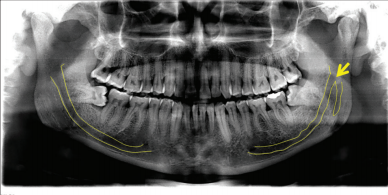
Introduction: Dental surgeries involving structures adjacent to the mandibular canal require greater knowledge of the intraosseous path, anatomical structure, and its variables, reducing the risk of injuries to this region. This research aimed to verify on the panoramic radiographs the anatomical characteristics of the mandibular canal, as well as to analyze and classify its pathways.
Methods: The classification of anatomical variations of the mandibular canal was divided into four types: Class A (inferior direction); Class B (mesial direction); Class C (alveolar direction); and Class D (retromolar direction). The sample consisted of 500 exams, 207 (41.4%) males and 293 (58.6%) females, with a mean age of 29.51 years.
Results: A prevalence of 30 anatomical variations of the mandibular canal was observed. The most prevalent classifications were Class B (43.6%) followed by Class C (23.1%) and D (33.3%). In no case was the presence of bifid canals classified as Class A. The anatomical variations of the mandibular canal appeared both unilaterally and bilaterally, in which unilaterally the prevalence was on the left side (50%), on the right side (20%), and bilaterally (30%).
Conclusions: According to the results obtained in this study, a prevalence of 6% of bifid mandibular canals was found. The most prevalent types of the bifid canal were Class B and Class D, and the highest occurrence of bifid mandibular canals was left unilateral. It is concluded that the appearance of mandibular canal anatomical variations in panoramic radiographs is frequent and that additional care must be taken to approach the region.
Downloads
Downloads
Published
License
Copyright (c) 2020 George Borja de Freitas, Pierre Gomes de Morais Silva, Jalber Almeida dos Santos, Luiz Roberto Coutinho Manhães Júnior, Paula Bernardon

This work is licensed under a Creative Commons Attribution 4.0 International License.
How to Cite