Effects of positional coracohumeral ligament stretching on the size of calcium deposits in adhesive capsulitis
DOI:
https://doi.org/10.17532/jhsci.2020.840Keywords:
Adhesive capsulitis, coracohumeral ligament, calcification, stretching, positional stretch, musculoskeletal ultrasoundAbstract
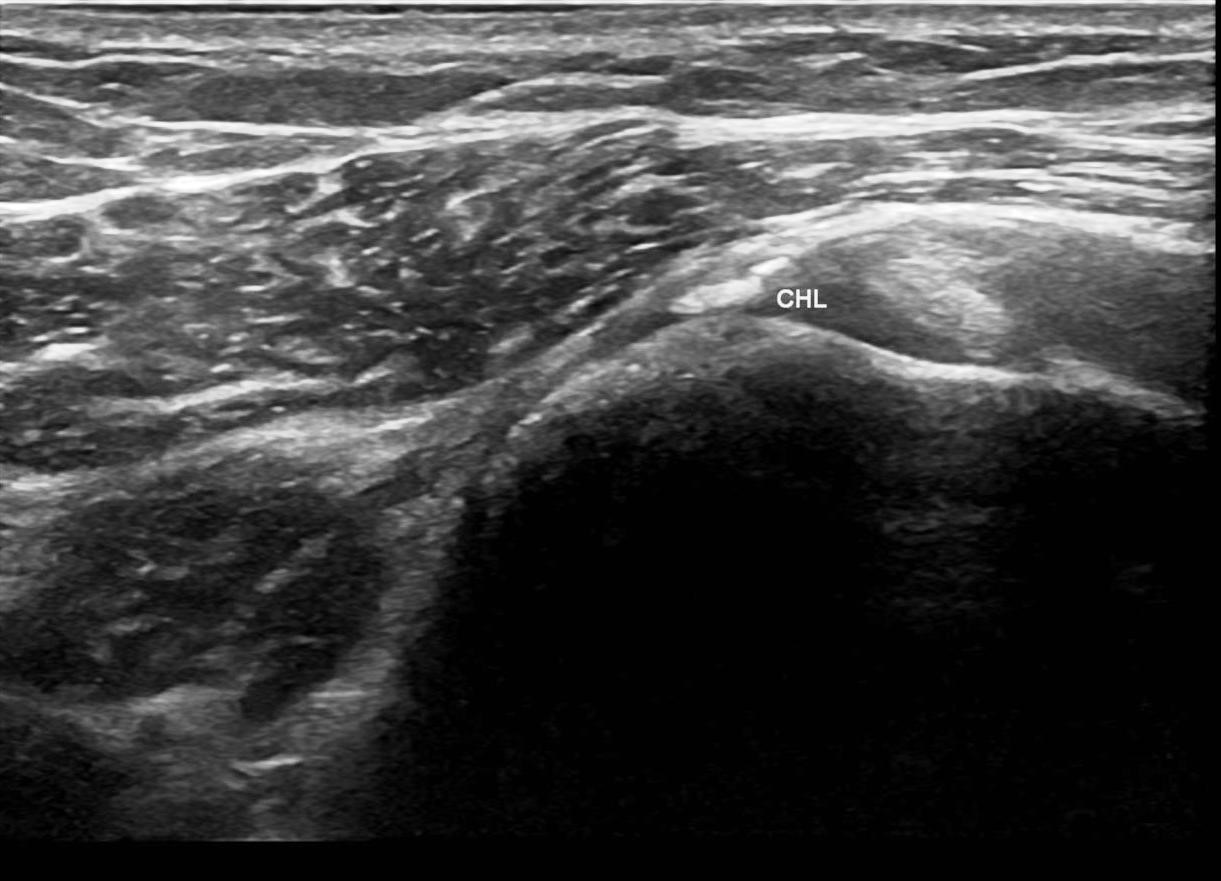
Adhesive capsulitis is a painful condition of unknown etiology with restriction of active and passive movements of the glenohumeral joint. The condition is a result of inflammation, adherence, and swelling in the lining of the shoulder joint capsule and its associated ligaments, causing resultant contracture of the capsule. We describe a patient with calcified and thickened coracohumeral ligament with adhesive capsulitis and diabetes mellitus.
Downloads
Download data is not yet available.
Downloads
Published
15.04.2020
How to Cite
1.
Effects of positional coracohumeral ligament stretching on the size of calcium deposits in adhesive capsulitis. JHSCI [Internet]. 2020 Apr. 15 [cited 2026 Jul. 25];10(1):99-102. Available from: https://www.jhsci.ba/ojs/index.php/jhsci/article/view/840