Vestibular bone thickness of the mandible in relation to the mandibular canal in a population from Bosnia and Herzegovina
DOI:
https://doi.org/10.17532/jhsci.2021.1293Keywords:
Cone-beam computed tomography, mandibular canal, vestibular thicknessAbstract
Introduction: Dental implantology is the branch of dentistry that is gaining greater significance because a larger number of patients come with requests of implant placements. During dental implant placements, with patients with whom operation is carried out in the mandible, very frequently nervus alveolaris inferior can be injured. The nerve injury may occur during the implant placement, but the nerve may also be injured in case of harvesting of intraoral bone graft. During the bone graft harvesting, but also during any other procedure in the dentistry that entails working on vestibular side of corpus of the mandible, in order not to injure the nervus alveolaris inferior, it is important to familiarize oneself with the distance of the nerve from the outer vestibular cortex of the mandible. The objective of the study was to assess the vestibular bone thickness of the mandible in relation to the mandibular canal with the help of analysis of cone-beam computed tomography (CBCT) images.
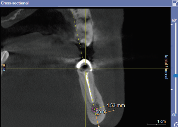
Methods: It was accessed the database of CBCT images taken at the School of Dental Medicine at the University of Sarajevo, where out of 700 reviewed CBCT images, an analysis of 322 CBCT images was conducted that satisfied inclusion criteria of the study. CBCT images were taken using of ORTHOPHOS SLX imaging unit. The measurement was conducted by Sidexis program on cross-section of CBCT image. The measurement of vestibular bone thickness was performed, by measuring the distance from the lateral wall of the mandibular canal to buccal mandibular compact bone, in the region of the second premolar, of the first and the second molar.
Results: There were statistically significant differences in vestibular bone thickness between men and women on both sides in the region of the second premolar (p < 0.001) and first molar (p = 0.016 right, p = 0.018 left). T-test demonstrated no statistically significant difference in the vestibular bone thickens between men and women on either side in the case of vestibular bone thickness of the center of the second molar (p = 0.397 right, p = 0.743 left).
Conclusion: Values of vestibular thickness of the mandible are larger with men than with women in all measuring points; however, statistically more significant differences between genders have been detected in the second premolar and center of the first molar.
Downloads
Downloads
Published
License
Copyright (c) 2021 Ajanović Muhamed, Selma Tosum Pošković, Alma Kamber-Ćesir, Edita Redžović, Mirsad Kacila, Karlo Kožul

This work is licensed under a Creative Commons Attribution 4.0 International License.
How to Cite