A comparison of erect weight-bearing and non-weight-bearing radiography of the cervical spine in non-trauma patients
DOI:
https://doi.org/10.17532/jhsci.2021.1261Keywords:
Cervical spine, radiography, computed radiography, digital radiography, cervicothoracic junction, weight-bearing techniqueAbstract
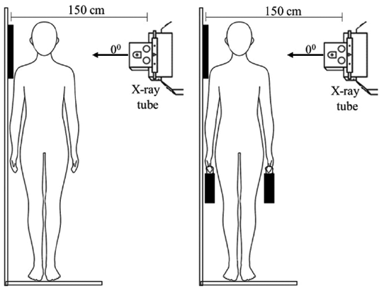
Introduction: Various positioning techniques are utilized to enhance the visualization of lower cervical vertebrae on lateral radiographs. However, the effectiveness of these techniques still remains unclear. This study was conducted to determine the effect of the weight-bearing (WB) technique in visualizing lower cervical vertebrae and cervicothoracic junction (C7-T1) on standing lateral cervical radiographs of adult non-trauma patients. The study was conducted using both computed radiography (CR) and digital radiography (DR) systems.
Methods: Forty-four CR (29 WB and 15 non-WB – NWB) and 61 DR (26 WB and 35 NWB) lateral C-spine radiographs were prospectively evaluated to assess the visible number of cervical vertebral bodies and C7-T1 junction. The instructions given by the radiographer to the patient for the imaging procedure were also assessed on the Likert scale (very good, good, fair, poor, very poor).
Results: There was no significant difference (p > 0.05) in the visualization of the number of vertebral bodies between the two techniques of WB and NWB for CR or DR. Further, no significant relationship (p > 0.05) was observed between the WB technique and the visualization of C7-T1 junction in DR systems. However, a significant difference was identified for CR (p = 0.012). The instruction given to the patient significantly correlated with the visibility of the lower C-spine region within each group of WB and NWB in both imaging systems.
Conclusions: The visibility of the number of vertebral bodies in the lower C-spine region in either CR or DR systems did not demonstrate any enhancement with the WB technique. Regardless of the imaging system or techniques used, adequate instructions given to the patient before and during the imaging procedure of C-spine lateral radiography demonstrated a significant improvement in visualizing the lower C-spine region. In this preliminary study, the application of erect WB radiography technique in evaluating the lower cervical region of adult non-trauma patients gives limited advantage.
Downloads
Downloads
Published
License
Copyright (c) 2021 Bimali S. Weerakoon*, Nimali N. Karunaratne, Winitha S. Jayasundara

This work is licensed under a Creative Commons Attribution 4.0 International License.
How to Cite