Computed tomography simulator conversion curve dependence on scan parameters and phantom dimension
DOI:
https://doi.org/10.17532/jhsci.2020.1085Keywords:
Computed tomography simulator, computed tomography-relative electron density, heterogeneous phantom, Hounsfield units, relative electron densityAbstract
Introduction: Using computed tomography (CT) and treatment planning systems (TPS) in radiotherapy, due to the difference in photon beam energy on CT and linear accelerator, it is necessary to convert Hounsfield units (HU) to relative electron density (RED) values. The aim of this dosimetric study was to determine whether there is a significant effect of potential in the CT tube, field of view size (FOV), and phantom dimensions on the CT conversion curve CT-RED. The second aim is whether there are significant differences between the CT-RED obtained by the Computerized Imaging Reference Systems (CIRS) Thorax 002LFC phantom and the “reference” curve in the TPS, obtained by the CIRS 062M pelvis phantom, at the same CT conditions.
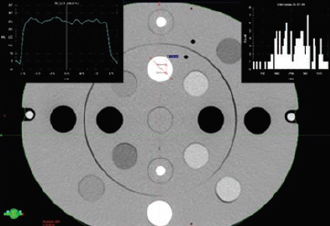
Methods: Heterogeneous CIRS 062M and CIRS Thorax 002LFC phantoms were used, which anatomically and dimensionally represent the human pelvis, head, and thorax, with a set of known RED inserts. They were scanned on a CT LightSpeed GE simulator and obtained CT-RED.
Results: The high voltage in the CT tube had a significant effect on the HU (t = 10.72, p < 0.001) for RED values >1.1, while FOV as a parameter did not show statistical significance for the 062M pelvis phantom. Comparing the slopes (062M pelvis and head) of the CT-RED for RED ≥ 1.1, the obtained value is t = 1.404 (p = 0.163). In the case of a 062M pelvis and a 002LFC phantom, we have seen a difference in RED values (for the same HU value) of 5 % in the RED region ≥ 1.1 (bone).
Conclusion: Patients should be imaged on a CT simulator only at the potential of the CT tube on which the conversion curve was recorded. The influence of the FOV and scanned phantom dimensions is not statistically significant on the appearance of the calibration curve (RED ≥ 1.1).
Downloads
Downloads
Published
License
Copyright (c) 2020 Goran Kolarević, Dražan Jaroš, Bojan Pavičar, Tatjana Ignjić, Aleksandar Kostovski, Goran Marošević, Branko Predojević, Dragoljub Mirjanić

This work is licensed under a Creative Commons Attribution 4.0 International License.
How to Cite